Which structure is highlighted and indicated by the leader line? Multiple Choice Posterior/dorsal horn o Anterior/ventral horn O Lateral horn 8. R 7R Which structure is highlighted and indicated by the leader line? Multiple Choice Lacrimal O Palatine O Ethmoid Sphenoid
The Correct Answer and Explanation is :
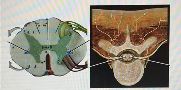
The highlighted structure indicated by the leader line in the provided image is the posterior (dorsal) horn of the spinal cord. This structure is one of the three grey columns of the spinal cord, along with the anterior (ventral) horn and lateral horn. The posterior horn is located dorsally and is involved in processing sensory information.
Anatomy and Function of the Posterior Horn:
The posterior horn, also known as the dorsal horn, is a prominent ridge of grey matter in the spinal cord. It extends to the surface of the spinal cord and is subdivided into six layers, termed Rexed laminae I-VI, based on the type of sensory information they process. These layers include:
- Lamina I: Marginal nucleus of the spinal cord
- Lamina II: Substantia gelatinosa of Rolando
- Laminae III and IV: Nucleus proprius
- Lamina V: Spinal lamina V, the neck of the posterior horn
- Lamina VI: Spinal lamina VI, the base of the posterior horn
The primary function of the posterior horn is to process and integrate sensory information from the peripheral nervous system. It receives inputs from primary afferent fibers and modulatory systems, projecting to higher brain centers and motor neurons. The dorsal horn circuitry is involved in various aspects of sensory processing, including discrimination, integration, and modulation of nociceptive (pain) and non-nociceptive signals. Dysfunction of the dorsal horn circuitry has been implicated in chronic pain conditions and other neurological disorders.
Anatomy and Function of the Palatine Bone:
The palatine bones are two irregular bones of the facial skeleton, located at the back of the nasal cavity between the maxilla and the pterygoid process of the sphenoid bone. They contribute to the walls of three cavities: the floor and lateral walls of the nasal cavity, the roof of the mouth, and the floor of the orbits. Each palatine bone consists of a horizontal plate, a perpendicular plate, and three projecting processes—the pyramidal process, which is directed backward and lateral from the junction of the two parts, and the orbital and sphenoidal processes, which surmount the vertical part. The two horizontal plates form the posterior part of the hard palate and the floor of the nasal cavity; anteriorly, they join with the maxillae. The palatine bones articulate with six bones: the sphenoid, ethmoid, maxilla, inferior nasal concha, vomer, and opposite palatine. They also contain foramina that transmit nerves and blood vessels to the region, including the greater and lesser palatine foramina.
In summary, the posterior horn of the spinal cord plays a crucial role in sensory processing, while the palatine bones are integral components of the facial skeleton, contributing to the structure of the nasal cavity, oral cavity, and orbit.