Correctly Label The Following Microscopic Anatomy Of The Hepatic Sinusoid. Erythrocytes In Sinusoid Fenestration MUUT Hepatic Macrophage Erythrocytes In Sinusoid Hepatocyte Central Vein Armyாராராராம் Endothelial Cells (UNION MINUS Und Sinusoid Bile Canaliculi Sinusoid Bile Ductule
The correct answer and explanation is:
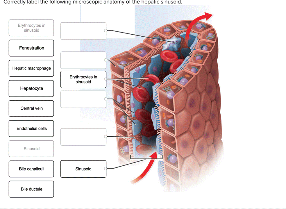
Correctly Labeled Microscopic Anatomy of the Hepatic Sinusoid:
- Erythrocytes in Sinusoid: Red blood cells found within the sinusoidal space.
- Fenestration: Small openings in the endothelial lining of the sinusoid allowing exchange between blood and liver cells.
- Hepatic Macrophage (Kupffer Cells): Specialized immune cells in the sinusoid responsible for filtering debris and pathogens from the blood.
- Hepatocyte: Liver cells involved in metabolism, detoxification, and bile production.
- Central Vein: The vein at the center of a liver lobule that collects blood from sinusoids.
- Endothelial Cells: Cells lining the walls of the sinusoid, forming the barrier between blood and liver tissue.
- Sinusoid: Specialized capillary in the liver where blood flows for filtration and nutrient exchange.
- Bile Canaliculi: Small ducts between hepatocytes where bile is collected before being transported to bile ductules.
- Bile Ductule: A small duct carrying bile from bile canaliculi to larger bile ducts.
Explanation:
The hepatic sinusoid is a unique vascular structure in the liver, designed for efficient exchange of nutrients, toxins, and immune interactions between blood and liver tissue. Blood from the hepatic portal vein and hepatic artery mixes and flows through the sinusoids, which are lined with fenestrated endothelial cells. These fenestrations facilitate the transfer of molecules between the blood and the space of Disse, where hepatocytes are located.
Kupffer cells, the liver’s resident macrophages, are attached to the endothelial lining and play a crucial role in immune surveillance and clearing debris. Erythrocytes flow within the sinusoidal lumen, carrying oxygen and nutrients.
Hepatocytes, arranged in plates, perform vital functions such as producing bile, metabolizing drugs, and synthesizing proteins. Bile produced by hepatocytes is secreted into bile canaliculi, small channels that converge into bile ductules, eventually leading to bile ducts.
At the center of each liver lobule is the central vein, where processed blood exits the liver. This structure is key to the liver’s ability to filter and detoxify blood while producing bile for digestion.