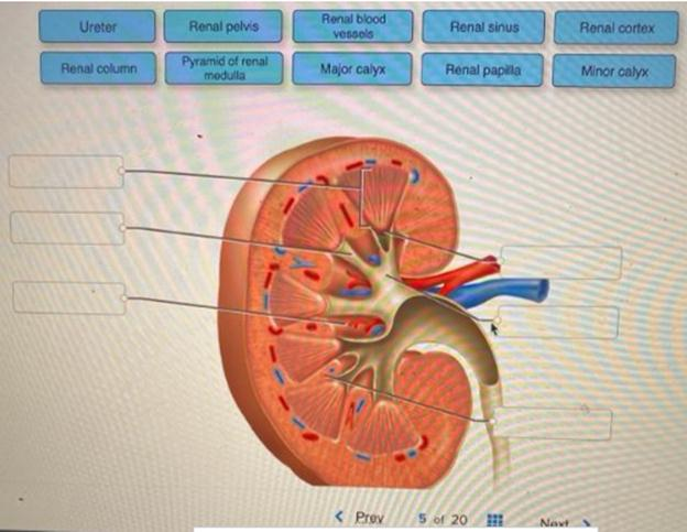
Correctly label the following anatomical parts of a kidney. __________________________________________________________________________________________________________________________________________________________________________________________________________________________________________
The correct answer and explanation is:
iturn0image1turn0image2turn0image5turn0image7The kidneys are vital organs responsible for filtering waste products, balancing electrolytes, and regulating blood pressure. Understanding their anatomy is crucial for comprehending their function. Below is a labeled diagram of the kidney, followed by detailed explanations of each anatomical part.
Labeled Diagram of the Kidney:
Anatomical Parts and Their Functions:
- Renal Capsule: A tough, fibrous layer surrounding the kidney, providing protection against trauma and infection.
- Renal Cortex: The outer region of the kidney beneath the capsule, containing the renal corpuscles and portions of the nephrons. It’s essential for filtering blood and initiating urine formation.
- Renal Medulla: The inner region, organized into cone-shaped structures called renal pyramids. It plays a crucial role in concentrating urine and maintaining water and salt balance.
- Renal Pyramids: Triangular structures within the medulla containing nephron loops and collecting ducts. They transport urine from the cortical areas to the calyces.
- Renal Columns: Extensions of cortical tissue between the pyramids, providing structural support and housing blood vessels.
- Renal Papilla: The apex of each pyramid, projecting into a minor calyx. It channels urine from the pyramids into the minor calyces.
- Minor Calyx: A small chamber that collects urine from a renal papilla. Multiple minor calyces converge to form a major calyx.
- Major Calyx: Formed by the union of minor calyces, these structures channel urine into the renal pelvis.
- Renal Pelvis: A funnel-shaped cavity that collects urine from the major calyces and transmits it to the ureter.
- Ureter: A muscular tube that conveys urine from the renal pelvis to the urinary bladder.
- Renal Artery: Supplies oxygenated blood to the kidney, branching into smaller arterioles within the cortex.
- Renal Vein: Drains deoxygenated blood from the kidney, returning it to the inferior vena cava.
- Nephron: The functional unit of the kidney, each consisting of a renal corpuscle and a renal tubule. Nephrons filter blood, reabsorb essential substances, and secrete waste into the forming urine.
- Renal Corpuscle: Comprises the glomerulus and Bowman’s capsule. It’s the initial filtering component of the nephron.
- Glomerulus: A network of capillaries where blood filtration begins, allowing water and small solutes to pass while retaining larger molecules like proteins.
- Bowman’s Capsule: A cup-like sac surrounding the glomerulus, collecting the filtrate to pass into the renal tubule.
- Proximal Convoluted Tubule (PCT): The first segment of the renal tubule, responsible for reabsorbing nutrients, electrolytes, and water from the filtrate back into the bloodstream.
- Loop of Henle: A U-shaped segment of the nephron that concentrates urine and conserves water by creating a concentration gradient in the medulla.
- Distal Convoluted Tubule (DCT): Further adjusts the composition of urine by reabsorbing sodium and calcium while secreting potassium and hydrogen ions.
- Collecting Duct: Receives urine from multiple nephrons, finalizing urine concentration before transporting it to the renal papilla.
- Renal Hilum: The recessed area on the medial side of the kidney where the renal artery, vein, and ureter enter and exit.
Understanding these structures provides insight into the kidney’s role in maintaining homeostasis, filtering blood, and producing urine.