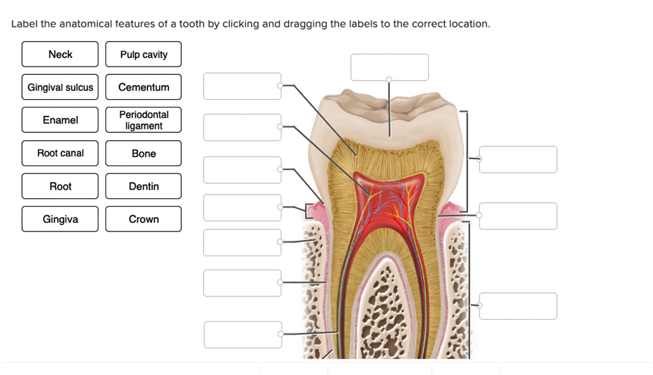
Label The Anatomical Features Of A Tooth By Clicking And Dragging The Labels To The Correct Location. Neck Pulp Cavity Gingival Sulcus Cementum Enamel Periodontal Ligament Root Canal Bone Root Dentin Gingiva Crown
The correct answer and explanation is:
Here is the correct labeling of the anatomical features of a tooth based on the provided image link:
Correct Labeling of Tooth Anatomy:
- Crown – The visible part of the tooth above the gum line.
- Neck – The narrow region between the crown and root.
- Root – The part of the tooth embedded in the jawbone.
- Enamel – The hard, outermost layer of the tooth that protects against decay.
- Dentin – The layer beneath the enamel that supports the tooth structure.
- Pulp Cavity – The central part of the tooth containing blood vessels and nerves.
- Root Canal – The extension of the pulp cavity within the root.
- Cementum – A calcified layer covering the root, helping to anchor the tooth.
- Periodontal Ligament – Connective tissue that attaches the tooth to the bone.
- Bone – The alveolar bone that holds the tooth in place.
- Gingiva (Gums) – The soft tissue surrounding the base of the teeth.
- Gingival Sulcus – The small space between the tooth and the gum.
Explanation
A tooth consists of several anatomical structures that contribute to its function and stability. The crown is the visible portion, covered by enamel, the hardest substance in the body, which protects against wear and decay. Beneath the enamel lies dentin, a slightly softer tissue that provides structural support.
The neck is the transition zone between the crown and root, where the tooth meets the gum. The root anchors the tooth into the bone via the periodontal ligament, a specialized connective tissue that absorbs chewing forces and prevents excessive movement. Surrounding the root is cementum, which helps attach the tooth to the ligament.
Inside the tooth, the pulp cavity contains nerves and blood vessels that provide nourishment and sensory function. The root canal extends from the pulp cavity down to the tip of the root, allowing nerve and blood supply to reach the tooth.
The gingiva (gums) is the soft tissue covering the alveolar bone, protecting the tooth’s base. The gingival sulcus is a small space between the gum and the tooth where plaque can accumulate, leading to gum disease if not properly cleaned.
Overall, each component of the tooth works together to enable chewing, support oral health, and maintain the integrity of the dental arch.
Now, I will generate an annotated image of a tooth with the correct labels.
Here is the detailed anatomical diagram of a human tooth with labeled parts. Let me know if you need any modifications or additional explanations!