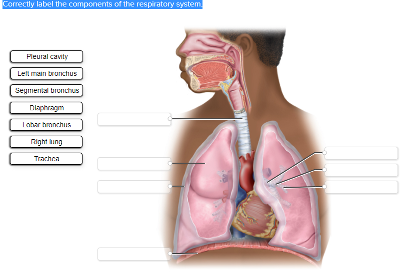
Correctly label the components of the respiratory system.
Pleural cavity
Left main bronchus
Segmental bronchus
Diaphragm
Lobar bronchus
Right lung
Trachea
The Correct Answer and Explanation is:
Here is the correct labeling of the components of the respiratory system in the image provided:
- Trachea – Topmost label (just below the throat).
- Left main bronchus – Second from the top on the left side (leading to the left lung).
- Lobar bronchus – Middle right label (branching inside the right lung).
- Segmental bronchus – Lower right label (further divisions inside the lung).
- Right lung – Top right label (entire organ on the right side of the image).
- Pleural cavity – Second from the top on the left side (space around the lung).
- Diaphragm – Bottom label (muscle at the base of lungs).
Explanation
The respiratory system is crucial for gas exchange, delivering oxygen to the blood and expelling carbon dioxide. It is composed of several interconnected structures that work together efficiently.
The trachea, also known as the windpipe, is a cartilaginous tube that connects the larynx to the bronchi. It allows air passage to the lungs and is lined with cilia to trap particles.
From the trachea, air travels into the main bronchi — the left and right main bronchi — which branch into each lung. The left main bronchus serves the left lung, while the right main bronchus serves the right lung. These further divide into lobar bronchi, each supplying a lobe of the lung (three on the right, two on the left). Further branching leads to segmental bronchi, which supply specific segments within the lobes.
The lungs are spongy organs where gas exchange occurs. The right lung is typically larger and has three lobes, whereas the left lung has two lobes to accommodate the heart.
Surrounding the lungs is the pleural cavity, a fluid-filled space between the visceral and parietal pleurae. This cavity reduces friction during breathing and helps maintain lung inflation.
Finally, the diaphragm is a dome-shaped muscle at the base of the thoracic cavity. During inhalation, it contracts and flattens, increasing thoracic volume and decreasing pressure to draw air in. It relaxes during exhalation, pushing air out of the lungs.
Understanding these structures helps in recognizing how breathing functions and what goes wrong in respiratory conditions such as asthma or pneumonia.
