On the following diagram of the cell cycle, label: Interphase, G1, S, G2, M Phase, Cytokinesis, metaphase, prophase, telophase, anaphase.
The Correct Answer and Explanation is:
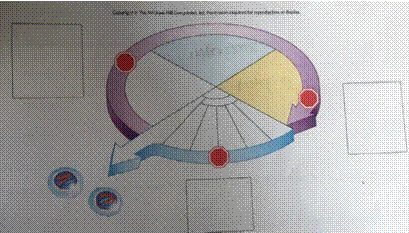
Based on the diagram you provided, which shows the cell cycle as a circular flow chart with distinct segments, here’s how the phases should be correctly labeled:
Labels for the Cell Cycle Diagram:
- Interphase – This includes G1, S, and G2 phases; it takes up the largest portion of the cycle (purple and blue regions combined).
- G1 Phase (Gap 1) – First section of interphase (light purple).
- S Phase (Synthesis) – Next segment where DNA is replicated (light blue).
- G2 Phase (Gap 2) – Final part of interphase (yellow region before mitosis).
- M Phase (Mitosis) – The phase where cell division occurs (entire arrow section).
- Cytokinesis – At the end of M phase, marked as the final split (arrowhead).
- Prophase – First part of mitosis (first segment in the M phase).
- Metaphase – Chromosomes align (second segment).
- Anaphase – Chromatids pull apart (third segment).
- Telophase – Final stage of mitosis before cytokinesis (last segment of M phase).
Explanation
The cell cycle is a regulated series of stages that cells go through to grow and divide. It has two major phases: Interphase and M phase (mitosis).
Interphase is the longest phase and consists of:
- G1 phase (Gap 1): The cell grows, synthesizes proteins, and carries out normal functions.
- S phase (Synthesis): DNA replication occurs, ensuring each daughter cell receives an identical set of chromosomes.
- G2 phase (Gap 2): The cell prepares for division, making proteins and organelles needed for mitosis.
After interphase, the cell enters the M phase, which includes both mitosis (division of the nucleus) and cytokinesis (division of the cytoplasm).
Mitosis is subdivided into four stages:
- Prophase: Chromatin condenses into visible chromosomes, and the nuclear envelope begins to break down.
- Metaphase: Chromosomes align at the cell’s equator.
- Anaphase: Sister chromatids are pulled apart to opposite poles by spindle fibers.
- Telophase: Chromosomes decondense, and nuclear envelopes re-form around the two nuclei.
Following mitosis, cytokinesis divides the cytoplasm, resulting in two genetically identical daughter cells. In animal cells, this involves a cleavage furrow that pinches the cell in two.
Understanding the cell cycle is crucial in fields like cancer research, where unregulated cell division plays a key role in disease progression.
Based on the diagram you provided, which shows the cell cycle as a circular flow chart with distinct segments, here’s how the phases should be correctly labeled:
.