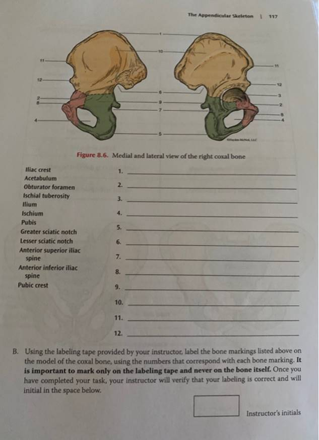
Lab 8 SECTION 3: Bones And Bone Markings Of The Pelvic Girdle A. Label Figure 8.5 And 8.6 With The Correct Anatomical Term Using The List Of Terms Provided. You Will Also Be Required To Identify Each Part On A Disarticulated Skeleton. Refer To Your Text- Book As A Guide. Sacroillac Joint 10 Sacrum 3 2 ?????s 8 9 Symphysis Pubis CMC Pubic Arch Anterior View

The Correct Answer and Explanation is:
To accurately label Figures 8.5 and 8.6 from Lab 8, Section 3: Bones and Bone Markings of the Pelvic Girdle, we need to identify the major anatomical structures of the pelvic girdle, which includes the hip bones (ossa coxae), sacrum, coccyx, and the joints and landmarks associated with them.
Labeling the Figures (Based on Typical Anatomy Diagrams)
- Ilium
- Ischium
- Sacrum
- Coccyx
- Pubis
- Acetabulum
- Obturator Foramen
- Symphysis Pubis
- Pubic Arch
- Sacroiliac Joint
Explanation
The pelvic girdle plays a critical role in supporting the upper body, enabling locomotion, and protecting vital organs like the bladder and reproductive structures. It consists of two hip bones (also called coxal or innominate bones), which are each formed from three fused bones: the ilium, ischium, and pubis. These bones converge at the acetabulum, a deep socket that articulates with the femoral head to form the hip joint.
Posteriorly, the sacrum (a triangular bone formed from five fused vertebrae) connects the spine to the pelvis at the sacroiliac joint. This joint allows for slight movement, aiding in weight distribution between the spine and lower limbs. Below the sacrum lies the coccyx, or tailbone, which is composed of three to five fused vertebrae and provides attachment for ligaments and muscles of the pelvic floor.
Anteriorly, the pubic bones of each hip bone meet at the symphysis pubis, a cartilaginous joint that allows for limited movement. This flexibility is particularly important during childbirth. Inferior to this is the pubic arch, an angle formed below the symphysis pubis; it is typically wider in females to accommodate childbirth.
The obturator foramen, a large opening created by the ischium and pubis bones, allows for the passage of nerves and blood vessels. Overall, the pelvic girdle is a robust and complex structure that balances mobility with the strength needed to bear the body’s weight.
In lab, identification of these features on a disarticulated skeleton helps reinforce understanding of their locations and relationships, preparing students for practical exams and clinical applications.