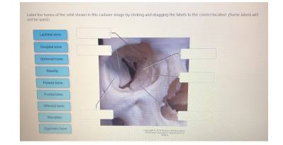
Label the bones of the orbit shown in this cadaver image by clicking and dragging the labels to the correct location. (Some labels will not be used.)
Lacrimal bone
Occipital bone
Sphenoid bone
Maxilla
Parietal bone
Frontal bone
Ethmoid bone
Mandible
Zygomatic bone
The Correct Answer and Explanation is:
Based on the image of the cadaver dissection showing the bones of the orbit (eye socket), the correctly labeled bones are as follows:
Correct Labels Placement:
- Frontal bone – Topmost region of the orbit (superior margin).
- Sphenoid bone – Deep within the posterior wall of the orbit.
- Zygomatic bone – Lateral wall of the orbit (cheekbone area).
- Maxilla – Inferior margin of the orbit (forms part of the floor).
- Lacrimal bone – Small bone near the medial wall of the orbit (next to the nasal cavity).
- Ethmoid bone – Medial wall, posterior to the lacrimal bone.
Labels NOT used:
- Occipital bone – Located at the posterior of the skull, not part of the orbit.
- Parietal bone – Forms part of the upper sides of the cranium, not involved in the orbit.
- Mandible – The lower jawbone, not connected to the orbit.
Explanation
The bony orbit is a pyramidal structure that houses the eye and associated structures. It is composed of seven bones that form the margins and walls of the orbit. Understanding the anatomical configuration of these bones is critical for medical students, particularly in fields like ophthalmology, maxillofacial surgery, and neurology.
- Frontal bone forms the superior border of the orbit and contributes to the roof. It also helps protect the eye from trauma.
- Sphenoid bone, specifically the lesser and greater wings, forms a major portion of the posterior orbital wall and contains the optic canal through which the optic nerve passes.
- Zygomatic bone contributes to the lateral wall and part of the floor. It is also a key structural element of the cheek.
- Maxilla, the upper jaw bone, forms the floor of the orbit and contains the infraorbital foramen for blood vessels and nerves.
- Lacrimal bone is one of the smallest and is situated in the anterior part of the medial orbital wall. It forms part of the nasolacrimal duct system, aiding tear drainage.
- Ethmoid bone lies posterior to the lacrimal bone and forms a significant portion of the medial wall of the orbit. It also contains the ethmoidal air cells (sinuses).
Understanding the arrangement of these bones is essential for diagnosing fractures, surgical planning, and understanding the spread of infections or tumors in the orbital region.