Label the following photos: Sheep Heart Dissection
Anterior View: Right side of the heart
Left side of the heart
Right and left auricles (appendages)
Apex
Pulmonary trunk
Aorta
Posterior View:
interventricular sulcus
Pulmonary veins
Vena cava
Interior View:
Left atrium
Left ventricle
Right atrium
Right ventricle
Interventricular septum
Bicuspid valve
Tricuspid valve
Chordae tendineae
Papillary muscles
The Correct Answer and Explanation is:
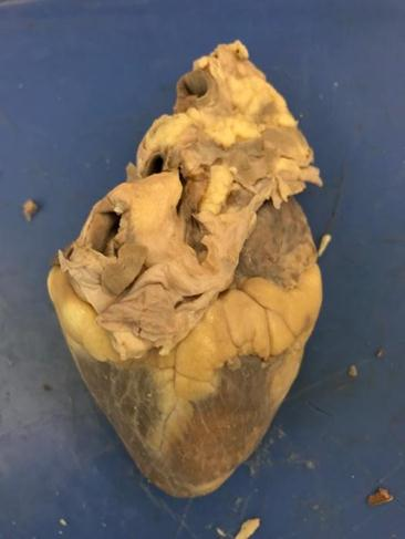
This image shows an external anterior view of a sheep heart. Here are the identified and labeled structures based on the anatomical features visible:
Anterior View Labels:
- Right Side of the Heart – On the left side of the image (remember, anatomical right is opposite of viewer’s left).
- Left Side of the Heart – On the right side of the image.
- Right and Left Auricles (Appendages) – Flap-like extensions near the top; the wrinkled, flap-looking structures are the auricles.
- Apex – The pointed bottom of the heart (visible at the bottom).
- Pulmonary Trunk – The large vessel coming out from the top, leftward of the image; this carries blood from the right ventricle to the lungs.
- Aorta – Slightly posterior to the pulmonary trunk, this is the thick-walled vessel that arches and carries blood from the left ventricle to the body.
Posterior View (Not visible in this photo):
- Interventricular Sulcus
- Pulmonary Veins
- Vena Cava
These structures are located on the back side of the heart and cannot be seen in this anterior view.
Interior View (Not visible in this photo):
- Left Atrium
- Left Ventricle
- Right Atrium
- Right Ventricle
- Interventricular Septum
- Bicuspid Valve
- Tricuspid Valve
- Chordae Tendineae
- Papillary Muscles
These structures are inside the heart and require dissection or cross-section to be visible.
Explanation
This image presents the anterior view of a preserved sheep heart, commonly used in anatomy and physiology studies due to its similarity in structure to the human heart. The heart apex, located at the bottom, points downward and to the left, which helps to determine anatomical orientation.
The right and left sides of the heart can be distinguished by the direction of the apex and the location of the large vessels. The right side (seen on the left in the image) includes the pulmonary trunk, which branches to send deoxygenated blood to the lungs. The left side (on the right of the image) includes the aorta, the thick-walled artery that distributes oxygenated blood to the body.
At the top, auricles (or appendages) of the atria are visible; these ear-like flaps increase the capacity of the atria. Their wrinkled appearance makes them distinguishable from the smoother surface of the ventricles.
Structures like the vena cava, pulmonary veins, and internal chambers (atria and ventricles), as well as valves such as the tricuspid, bicuspid, and chordae tendineae, are found internally or on the posterior and thus are not visible from this angle. Dissecting the heart or rotating it to a posterior view would reveal those features.
Understanding the orientation and surface features of the heart is vital for dissecting and identifying structures accurately. This anterior view forms the basis for exploring internal anatomy and understanding blood flow through the heart.