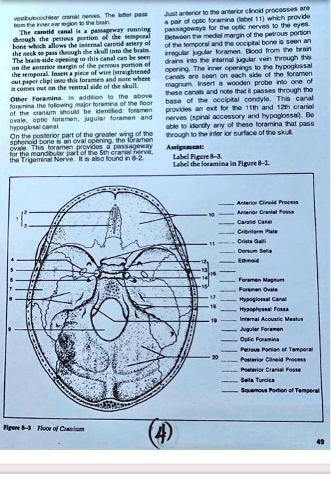
vestibulocochlear cranial nerves. The latter pass from the inner ear region to the brain. The carotid canal is a passageway running through the petrous portion of the temporal bone which allows the internal carotid artery of the neck to pass through the skull into the brain. The brain-side opening to this canal can be seen on the anterior margin of the petrous portion of the temporal. Insert a piece of wire (straightened out paper clip) into this foramen and note where it comes out on the ventral side of the skull. Other Foramina. In addition to the above foramina the following major foramina of the floor of the cranium should be identified: foramen ovale, optic foramen, jugular foramen and hypoglosal canal. On the posterior part of the greater wing of the sphenoid bone is an oval opening, the foramen ovale. This foramen provides a passageway for the mandibular part of the 5th cranial nerve, the Trigeminal Nerve. It is also found in 8-2. Just anterior to the anterior clinoid processes are a pair of optic foramina (label 11) which provide passageways for the optic nerves to the eyes. Between the medial margin of the petrous portion of the temporal and the occipital bone is seen an irregular jugular foramen. Blood from the brain drains into the internal jugular vein through this opening. The inner openings to the hypoglossal canals are seen on each side of the foramen magnum. Insert a wooden probe into one of these canals and note that it passes through the base of the occipital condyle. This canal provides an exit for the 11th and 12th cranial nerves (spinal accessory and hypoglossal). Be able to identify any of these foramina that pass through to the inferior surface of the skull. Assignment: Label Figure 8-3. Label the foramina in Figure 8-2. Figure 8-3 Floor of Cranium Anterior Clinoid Process Anterior Cranial Fossa Carotid Canal Cribriform Plate Crista Galli Dorsum Sella Ethmoid Foramen Magnum Foramen Ovale Hypoglossal Canal Hypophyseal Fossa Internal Acoustic Meatus Jugular Foramen Optic Foramina Petrous Portion of Temporal Posterior Clinoid Process Posterior Cranial Fossa Sella Turcica Squamous Portion of Temporal
The Correct Answer and Explanation is:
Based on the anatomical features presented in the diagram and the provided list of terms, here is the correct labeling for Figure 8-3.
Correct Labels for Figure 8-3:
- Crista Galli
- Cribriform Plate
- Ethmoid
- Squamous Portion of Temporal
- Foramen Ovale
- Carotid Canal
- Petrous Portion of Temporal
- Jugular Foramen
- Foramen Magnum
- Anterior Cranial Fossa
- Optic Foramina
- Anterior Clinoid Process
- Sella Turcica
- Dorsum Sella
- Posterior Clinoid Process
- Hypophyseal Fossa
- Hypoglossal Canal
- Internal Acoustic Meatus
- Jugular Foramen
- Posterior Cranial Fossa
Explanation of the Floor of the Cranium
The floor of the cranium, viewed from above, reveals a complex and uneven surface divided into three distinct depressions, or fossae, which accommodate the different lobes of the brain. These are the anterior, middle, and posterior cranial fossae.
The Anterior Cranial Fossa (10) is the highest and most forward-located fossa, supporting the frontal lobes of the cerebrum. Its central feature is formed by the Ethmoid (3) bone, which includes the Crista Galli (1), a prominent vertical crest for the attachment of the falx cerebri, and the perforated Cribriform Plate (2) on either side, which allows passage for the olfactory nerves from the nasal cavity to the brain.
The butterfly-shaped middle cranial fossa houses the temporal lobes. Its central, elevated part is the Sella Turcica (13) (“Turkish saddle”) of the sphenoid bone. This structure includes the Hypophyseal Fossa (16), which cradles the pituitary gland, and is bordered by the Anterior (12) and Posterior (15) Clinoid Processes and the Dorsum Sella (14). Important openings in this fossa include the Optic Foramina (11) for the optic nerves, the Foramen Ovale (5) for the trigeminal nerve, and the internal opening of the Carotid Canal (6) for the internal carotid artery. The lateral walls of this fossa are formed by the Squamous Portion of the Temporal bone (4).
The Posterior Cranial Fossa (20) is the largest and deepest, containing the cerebellum, pons, and medulla oblongata. It is separated from the middle fossa by the dense Petrous Portion of the Temporal bone (7). Its most conspicuous feature is the Foramen Magnum (9), the large opening through which the spinal cord connects with the brainstem. Other vital openings include the Jugular Foramen (8, 19) for the internal jugular vein and cranial nerves IX, X, and XI; the Internal Acoustic Meatus (18) for cranial nerves VII and VIII; and the Hypoglossal Canal (17) for cranial nerve XII.
