Hcle Save & Exit Below are the features of the image of simple columnar epithelium using the terms provided: Connective tissue Nucleus Basement membrane Cytoplasm Goblet cells Microvilli Lumen
The Correct Answer and Explanation is:
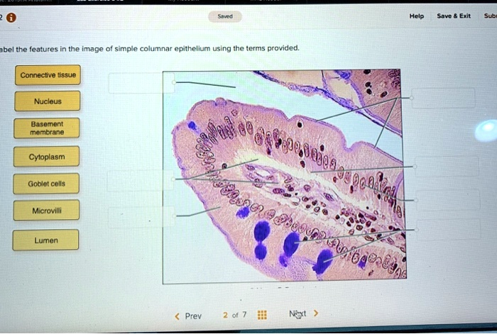
Here are the correct labels for the features of the simple columnar epithelium:
- Top-left pointer: Microvilli
- Second pointer from top-left: Goblet cells
- Third pointer from top-left: Connective tissue
- Top-right pointer: Lumen
- Second pointer from top-right: Nucleus
- Third pointer from top-right: Cytoplasm
- Bottom-right pointer: Basement membrane
Explanation
This image displays a micrograph of simple columnar epithelium, a tissue type commonly found lining the gastrointestinal tract, such as in the small intestine. This tissue is specialized for absorption and secretion.
The Lumen is the open space or cavity within a tubular organ, in this case, the intestine. This is where digested food, known as chyme, would be located.
The epithelial surface facing the lumen is covered in Microvilli. These are microscopic, finger-like projections of the cell membrane that form a structure called a brush border. Their primary function is to dramatically increase the surface area of the cell, which is crucial for maximizing the absorption of nutrients from the digested food.
Interspersed among the columnar absorptive cells are Goblet cells. These are specialized, mucus-secreting cells, easily identified by their flask-like shape and the accumulation of purple-staining mucin granules. The mucus they produce lubricates the intestinal lining, protecting it from mechanical abrasion and chemical damage from digestive enzymes.
Each columnar cell contains a Nucleus, a large organelle that houses the cell’s genetic material and controls its activities. In simple columnar epithelium, the nuclei are typically elongated and aligned in a row near the basal side of the cells. The material filling the rest of the cell is the Cytoplasm, which includes the cytosol and all other organelles where metabolic processes take place.
The entire epithelial layer is anchored to the underlying tissue by the Basement membrane. This is a thin, non-cellular layer of extracellular proteins that provides structural support and acts as a selective filter.
Deep to the basement membrane is the Connective tissue, specifically the lamina propria. This layer is rich in blood vessels, lymphatic vessels, and immune cells, providing vital metabolic support and defense for the overlying epithelium.