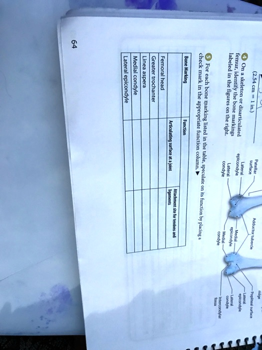
2.54 cm = 1 in.) 4 On a skeleton or disarticulated femur, identify the bone markings labeled in the figures on the right. ? For each bone marking listed in the table, speculate on its function by placing a check mark in the appropriate function column. Bone Marking Function Articulating surface at a joint Attachment site for tendons and ligaments Femoral head Greater trochanter Linea aspera Medial condyle Lateral epicondyle
The Correct Answer and Explanation is:
Here is the correct answer for the table, followed by a detailed explanation.
| Bone Marking | Articulating surface at a joint | Attachment site for tendons and ligaments |
| Femoral head | ✓ | |
| Greater trochanter | ✓ | |
| Linea aspera | ✓ | |
| Medial condyle | ✓ | |
| Lateral epicondyle | ✓ |
Explanation
The markings on a bone’s surface are not random; they directly relate to their specific physiological functions, primarily falling into two categories: forming joints or serving as attachment points for connective tissues.
The Femoral head is the large, rounded structure at the proximal end of the femur. Its smooth, spherical shape is designed to fit perfectly into the acetabulum of the pelvic bone, forming the hip joint. This ball-and-socket joint allows for a wide range of motion. Therefore, its primary function is to serve as an articulating surface.
In contrast, the Greater trochanter is a large, irregular prominence located on the lateral side of the femur’s proximal end. Its rough surface indicates that it is an attachment site for powerful muscles. Several muscles that move the thigh, including the gluteus medius and gluteus minimus, attach here via their tendons.
Similarly, the Linea aspera is a long, vertical ridge on the posterior shaft of the femur. Its name translates to “rough line,” which accurately describes its function as a major attachment site for numerous muscles. The powerful adductor, hamstring, and quadriceps muscles of the thigh all have origins or insertions along this line.
At the distal end of the femur, the Medial condyle is one of two large, smooth knobs that form the knee joint. It articulates with the medial condyle of the tibia. Like the femoral head, its smooth cartilage-covered surface is essential for low-friction movement, specifically the flexion and extension of the leg at the knee.
Finally, the Lateral epicondyle is a smaller bony projection situated on the outer side of the femur, just above the lateral condyle. The prefix “epi” means “upon,” signifying its position relative to the articulating condyle. Epicondyles are not part of the joint’s articulating surface but instead serve as crucial anchor points. The lateral epicondyle is the attachment site for the lateral collateral ligament (LCL) of the knee, which provides stability to the joint.