Microvillus Plasma membrane Centrosome Microtubule Cilium Microfilament Microtubule Centrosome Plasma membrane Microvillus Microfilament
Label the structures of the plasma membrane and cytoskeleton. Microvillus Plasma membrane Centrosome Microtubule Cilium Microfilament Microtubule Centrosome Plasma membrane Microvillus Microfilament
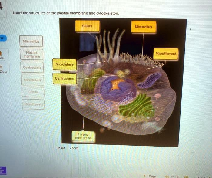
The Correct Answer and Explanation is:
Based on the diagram provided, here are the correct labels for the indicated structures:
- Cilium: The long, hair-like projection extending from the cell surface.
- Microvillus: One of the short, finger-like projections that increase the cell’s surface area.
- Microfilament: The thin protein fiber located just beneath the plasma membrane and forming the core of the microvilli.
- Microtubule: The thick, hollow tube-like protein filament within the cytoplasm.
- Centrosome: The organelle located near the nucleus that organizes microtubules.
- Plasma membrane: The outer boundary of the cell.
Explanation of the Plasma Membrane and Cytoskeleton
The provided diagram illustrates key components of an animal cell’s outer boundary and internal framework. The plasma membrane is the cell’s essential outer layer. It functions as a selective barrier, meticulously controlling which substances enter and leave the cell. This regulation is crucial for maintaining a stable internal environment, or homeostasis.
Internal to the plasma membrane is the cytoskeleton, a dynamic network of protein filaments that provides structural support, determines cell shape, and facilitates movement. The diagram highlights several cytoskeletal elements. Microfilaments are the thinnest of these filaments, composed of the protein actin. They form a supportive mesh just under the plasma membrane and are responsible for cell motility, changes in cell shape, and muscle contraction. As shown, they provide the structural core for microvilli, which are short, non-motile projections that dramatically increase the cell’s surface area for absorption.
Microtubules are the largest components of the cytoskeleton. These hollow tubes, made of tubulin protein, act like highways within the cell, providing tracks along which organelles and vesicles can move. They also play a critical role in cell division by forming the mitotic spindle. Microtubules are organized by the centrosome, which is the primary microtubule-organizing center in animal cells. The centrosome is typically located near the nucleus and contains a pair of centrioles.
Finally, some microtubules assemble into more complex structures like cilia. Cilia are long, hair-like projections that can move in a coordinated, wave-like fashion. Their function is either to propel the entire cell through a fluid or to move fluid and particles across the cell’s surface, as seen in the respiratory tract.