The Correct Answer and Explanation is:
The correct answer is ependymal cells.
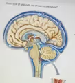
The glial cells prominently illustrated in this sagittal view of the brain are ependymal cells. This identification is based on their specific location and implied function as depicted in the diagram. Ependymal cells are a type of neuroglia, or glial cell, that form a simple cuboidal epithelial-like lining called the ependyma. This lining covers the inner surfaces of the brain’s ventricles, which are the fluid filled cavities, and the central canal of the spinal cord. In the figure, these cells are shown lining the large, interconnected spaces within the brain that are colored blue, representing the presence of cerebrospinal fluid (CSF).
The primary and most vital function of ependymal cells is the production and regulation of cerebrospinal fluid. Specialized clusters of these cells, combined with a network of capillaries, form a structure known as the choroid plexus. The reddish, tufted structures shown within the ventricles in the diagram represent this choroid plexus, the site where most CSF is synthesized. By filtering blood plasma, the ependymal cells of the choroid plexus secrete CSF into the ventricular system.
Furthermore, many ependymal cells possess cilia, which are small, hair like projections on their surface facing the ventricles. These cilia beat in a coordinated rhythm to help circulate the CSF throughout the brain and spinal cord. This circulation is critical for several reasons. CSF provides buoyancy, effectively reducing the brain’s weight and preventing it from being crushed under its own mass. It also acts as a crucial shock absorber, protecting the delicate neural tissue from mechanical injury. Finally, the CSF facilitates the transport of nutrients to the brain and the removal of metabolic waste products. The diagram’s emphasis on the ventricular system and the fluid it contains makes it clear that the illustrated glial cells are the ependymal cells responsible for these processes.