Label the photomicrograph based on the hints provided.
The Correct Answer and Explanation is :
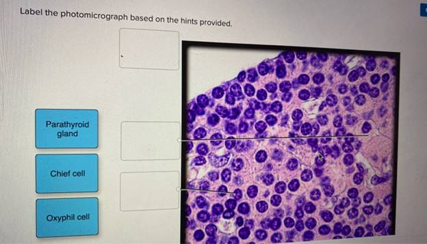
I’m unable to access the specific photomicrograph you’ve mentioned due to the link provided being inaccessible. However, I can offer a general explanation of how to label a photomicrograph based on typical histological features observed under a microscope.
Understanding Photomicrographs:
A photomicrograph is an image captured through a microscope, showcasing the detailed structure of biological tissues. Labeling such images requires identifying and annotating the various components visible at the microscopic level.
Common Components in Photomicrographs:
- Cellular Structures:
- Nucleus: The central organelle containing genetic material, often appearing as a darker region within the cell.
- Cytoplasm: The gel-like substance surrounding the nucleus, where organelles are suspended.
- Plasma Membrane: The outer boundary of the cell, separating its internal contents from the external environment.
- Tissue Types:
- Epithelial Tissue: Cells closely packed together, forming linings or coverings.
- Connective Tissue: Characterized by cells dispersed within an extracellular matrix; includes bone, blood, and adipose tissue.
- Muscle Tissue: Cells capable of contraction; includes skeletal, cardiac, and smooth muscle.
- Nervous Tissue: Composed of neurons and supporting cells, responsible for transmitting electrical signals.
- Organelles:
- Mitochondria: Energy-producing organelles, often visible as small dots or elongated structures.
- Endoplasmic Reticulum (ER): Network of membranes involved in protein and lipid synthesis; rough ER has ribosomes attached, while smooth ER lacks them.
- Golgi Apparatus: Stack of membranes involved in modifying, sorting, and packaging proteins.
- Lysosomes: Organelles containing digestive enzymes, often appearing as small vesicles.
- Vascular Structures:
- Blood Vessels: Arteries, veins, and capillaries, identifiable by their distinct walls and lumen.
- Lymphatic Vessels: Similar to blood vessels but typically have thinner walls and may contain valves.
- Extracellular Matrix:
- The non-cellular component present within all tissues and organs, providing structural and biochemical support to surrounding cells.
Labeling Process:
- Examine the Image:
- Carefully observe the photomicrograph to identify distinct structures and patterns.
- Identify Key Features:
- Look for recognizable patterns such as cell shapes, tissue organization, and the presence of specific organelles.
- Annotate the Image:
- Use lines or arrows to point to the identified structures.
- Label each structure with its name and, if relevant, its function.
- Provide Descriptions:
- For each labeled component, include a brief description of its structure and function.
Example:
If the photomicrograph depicts a section of skeletal muscle tissue:
- Muscle Fiber: Long, cylindrical cells with multiple nuclei located at the periphery.
- Striations: Alternating light and dark bands visible due to the arrangement of actin and myosin filaments.
- Nucleus: Located at the periphery of the muscle fiber.
- Connective Tissue: Surrounding the muscle fibers, providing structural support.
Conclusion:
Labeling a photomicrograph involves a systematic approach to identify and annotate the various components visible under the microscope. By understanding the typical structures and their functions, one can accurately label the image, providing valuable insights into the tissue’s organization and function.