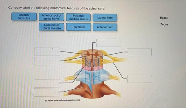
Correctly label the following anatomical features of the spinal cord. Anterior Anterior root of Posterior tuniculus spinal nerve Lateral horn median sulcus Reset Dura mater Zoom (dural sheath) Pia mater Anterior hom Spinal cord and meninge therack)
The Correct Answer and Explanation is :
iturn0image0turn0image2turn0image4turn0image8The spinal cord is a vital component of the central nervous system, serving as the primary conduit for neural signals between the brain and the body. Its structure is organized into several distinct anatomical features, each with specific functions.
Anterior Median Fissure: This is a deep groove running along the midline of the anterior (ventral) surface of the spinal cord. It partially divides the spinal cord into two symmetrical halves and serves as a landmark for orientation.
Posterior Median Sulcus: Located on the posterior (dorsal) surface, this shallow groove runs along the midline and, like the anterior median fissure, helps in identifying the spinal cord’s symmetry.
Anterior Horn: Also known as the ventral horn, this is the front portion of the gray matter in the spinal cord. It contains motor neurons that send signals to muscles, facilitating voluntary movement.
Posterior Horn: Referred to as the dorsal horn, this is the rear portion of the gray matter. It processes sensory information received from the body, playing a crucial role in sensation.
Lateral Horn: Present in certain segments of the spinal cord, particularly in the thoracic and upper lumbar regions, the lateral horn contains autonomic motor neurons that regulate involuntary functions such as heart rate and digestion.
Dura Mater: This is the outermost layer of the meninges, the protective membranes surrounding the spinal cord. It is a tough, fibrous layer that provides structural support and protection.
Arachnoid Mater: Situated between the dura mater and pia mater, the arachnoid mater is a web-like structure that contains the cerebrospinal fluid, cushioning the spinal cord and absorbing shocks.
Pia Mater: The innermost layer of the meninges, the pia mater is a delicate membrane that closely adheres to the surface of the spinal cord, following its contours and supplying it with blood vessels.
Spinal Nerve Roots: These are the initial segments of the spinal nerves, emerging from the spinal cord. Each spinal nerve is formed by the union of a dorsal (sensory) root and a ventral (motor) root.
Understanding these anatomical features is essential for comprehending how the spinal cord functions in transmitting neural signals, coordinating movement, and processing sensory information.