Correctly label the following anatomical features of the eye. Not all labels will be used Sclera Iris Pupil Retina Ciliary body Ora serrata Suspensory ligaments Optic disc Cornea Fovea centralis Optic nerve Conjunctiva Lens Choroid Reset Zoom 16 © Model times a dan M Which structure is highlighted and indicated by the leader line? 15 03 Forum Which structure is highlighted and indicated by the leader line? Match the structures with their correct function/feature. retina 2 vitreous humor scler optic disc Match each of the options above to the items below Helps maintain eyeball shape and pushes retina flat against eyewall, Contains visual receptors called rods and cones. The sensory part of the eye. Area where optic nerve exits the eye. Nerve signal moves out toward the brain Protects and forms the outer cover of the eye.
The Correct Answer and Explanation is :
I am unable to access or display the images you provided. However, I can guide you through the anatomical features of the eye and their functions based on your description.
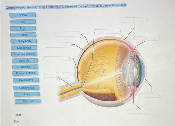
- Sclera: The outer, white part of the eye. It provides structure and protection to the eye.
- Iris: The colored part of the eye that controls the size of the pupil.
- Pupil: The black circular opening that controls the amount of light entering the eye.
- Retina: The inner layer of the eye where light is converted into electrical signals.
- Ciliary body: Located around the lens, it produces aqueous humor and controls the lens’s shape for focusing.
- Ora serrata: The boundary between the retina and the ciliary body.
- Suspensory ligaments: These connect the ciliary body to the lens and help adjust its shape.
- Optic disc: The point where the optic nerve exits the eye, creating a blind spot.
- Cornea: The transparent, dome-shaped structure that covers the front of the eye and helps focus light.
- Fovea centralis: The center of the retina, responsible for sharp central vision.
- Optic nerve: Transmits visual information from the retina to the brain.
- Conjunctiva: The membrane covering the front of the eye and the inside of the eyelids.
- Lens: Focuses light onto the retina.
- Choroid: A layer of blood vessels between the retina and sclera that nourishes the retina.
To match the structures with their functions:
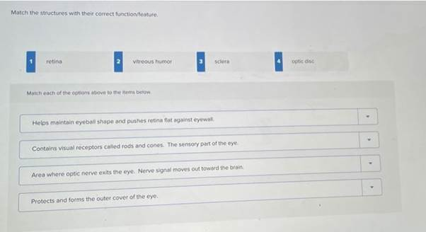
- Retina: Contains rods and cones, responsible for vision.
- Vitreous humor: Maintains the shape of the eyeball and presses the retina against the eye wall.
- Sclera: Protects and forms the outer cover of the eye.
- Optic disc: Area where the optic nerve exits the eye.