Use the list of terms provided to label the accompanying illustration of a skeletal muscle fiber; check off each term as you label it.
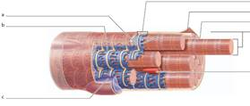
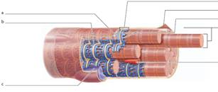
□ myofibrils
□ nucleus
□ sarcolemma
□ sarcoplasmic reticulum
□ T-tubule
□ terminal cisternae
□ triad
The Correct Answer and Explanation is :
A skeletal muscle fiber, or muscle cell, is a complex structure composed of various specialized components that facilitate muscle contraction. Here’s an overview of the key structures:
- Myofibrils: These are long, cylindrical organelles within the muscle fiber, composed of repeating units called sarcomeres. Sarcomeres contain the contractile proteins actin and myosin, which interact to produce muscle contraction through the sliding filament mechanism.
- Nucleus: Skeletal muscle fibers are multinucleated, meaning they contain multiple nuclei located just beneath the cell membrane (sarcolemma). These nuclei are essential for regulating gene expression and protein synthesis necessary for muscle function and adaptation.
- Sarcolemma: This is the plasma membrane of the muscle fiber. It encloses the cell’s contents and plays a crucial role in conducting electrical impulses (action potentials) that trigger muscle contraction.
- Sarcoplasmic Reticulum (SR): A specialized form of smooth endoplasmic reticulum that surrounds each myofibril. The SR stores calcium ions (Ca²⁺) and releases them into the sarcoplasm (muscle cell cytoplasm) in response to an action potential, initiating muscle contraction.
- T-Tubule (Transverse Tubule): These are invaginations of the sarcolemma that penetrate into the muscle fiber’s interior. T-tubules conduct action potentials from the cell surface to deeper regions, ensuring a uniform and rapid activation of the muscle fiber.
- Terminal Cisternae: Enlarged areas of the sarcoplasmic reticulum adjacent to the T-tubules. They work in conjunction with T-tubules to facilitate the release of calcium ions during muscle contraction.
- Triad: A structural formation consisting of a T-tubule flanked by two terminal cisternae. The triad is essential for coupling the electrical signal of an action potential with the release of calcium ions from the sarcoplasmic reticulum, a process known as excitation-contraction coupling.
Understanding these components is vital for comprehending how skeletal muscles contract. When an action potential travels along the sarcolemma and down the T-tubules, it triggers the terminal cisternae of the sarcoplasmic reticulum to release calcium ions into the sarcoplasm. The increase in calcium concentration allows actin and myosin within the myofibrils to interact, leading to sarcomere shortening and, consequently, muscle contraction. After contraction, calcium ions are pumped back into the sarcoplasmic reticulum, allowing the muscle to relax.
This intricate system ensures that muscle fibers contract efficiently and respond rapidly to neural stimuli, enabling precise and coordinated movements.