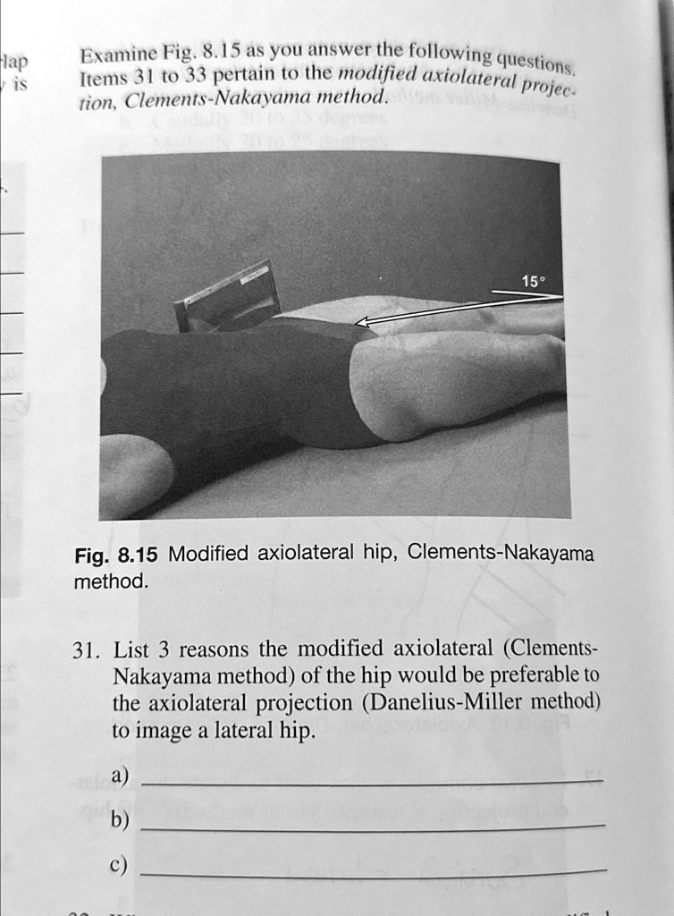
Examine Fig. 8.15 as you answer the following questions. Items 31 to 33 pertain to the modified axiolateral projection, Clements-Nakayama method. 15° Fig. 8.15 Modified axiolateral hip, Clements-Nakayama method. 31. List 3 reasons the modified axiolateral (Clements- Nakayama method) of the hip would be preferable to the axiolateral projection (Danelius-Miller method) to image a lateral hip. a) b) c)
The correct Answer and Explanation is:
Three reasons why the modified axiolateral (Clements-Nakayama) method is preferable to the axiolateral (Danelius-Miller) method for imaging a lateral hip:
- Suitable for patients who cannot elevate the unaffected leg – The Danelius-Miller method requires the patient to lift the unaffected limb out of the imaging field, which may not be possible in cases of severe trauma or limited mobility. The Clements-Nakayama method allows imaging without requiring the patient to move excessively.
- Minimizes distortion and magnification – The Clements-Nakayama method incorporates a 15-degree tilt of the image receptor and a matching angulation of the X-ray beam to optimize visualization of the femoral neck while reducing distortion compared to the traditional axiolateral technique.
- Less invasive for critically injured patients – Since it does not require extensive patient manipulation, this method is especially useful for imaging patients with hip fractures, post-surgical conditions, or other trauma-related injuries where movement might cause significant discomfort or worsen the condition.
Explanation
The modified axiolateral projection, known as the Clements-Nakayama method, is a valuable technique used in radiography for patients who cannot assume the standard positioning required for an axiolateral hip projection, such as the Danelius-Miller method. This modification is particularly beneficial for trauma patients, post-surgical individuals, and those with severe mobility limitations.
One of the key advantages of the Clements-Nakayama method is its ability to accommodate patients who are unable to elevate the unaffected limb, a requirement in the Danelius-Miller method. In cases of bilateral hip fractures or severe pain, lifting the leg may be impractical or impossible. By adjusting both the image receptor and the X-ray beam to a 15-degree tilt, this technique ensures that the femoral neck remains properly visualized while avoiding the interference of the contralateral limb.
Another advantage lies in the reduction of geometric distortion and magnification. Because the image receptor is slightly angled rather than remaining perfectly perpendicular to the X-ray beam, the Clements-Nakayama method enhances the clarity of the femoral neck while maintaining anatomical accuracy. This improved visualization is crucial for assessing fractures, bone integrity, and surgical implants.
Finally, minimizing movement in trauma patients is essential for preventing further injury and reducing discomfort. The Clements-Nakayama method eliminates the need for extensive repositioning, making it a preferred choice for individuals in critical conditions. By allowing radiologists to obtain clear and diagnostic images without excessive patient manipulation, this method enhances the efficiency and safety of hip imaging procedures.