Correctly label the following parts of a skeletal muscle fiber. A band Sarcoplasmic reticulum Myofilaments Sarcoplasm Myofibril I band Sarcolemma Z disc Mitochondria Muscle fiber Reset Zoom
The Correct Answer and Explanation is:
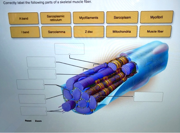
The correct labeling of the parts of a skeletal muscle fiber is as follows:
- A band – The region of the sarcomere that contains thick filaments, appearing as a dark band.
- Sarcoplasmic reticulum – A specialized network surrounding myofibrils, responsible for storing and releasing calcium ions.
- Myofilaments – The actin and myosin filaments that form the contractile structures within myofibrils.
- Sarcoplasm – The cytoplasm of a muscle fiber, containing essential components such as mitochondria and enzymes.
- Myofibril – A cylindrical structure within the muscle fiber composed of repeating sarcomeres.
- I band – The light band adjacent to the A band, containing only thin filaments.
- Sarcolemma – The plasma membrane that encloses the muscle fiber and facilitates signal transmission.
- Z disc – The structural boundary of sarcomeres, anchoring the thin filaments.
- Mitochondria – Organelles responsible for generating ATP, providing energy for muscle contraction.
- Muscle fiber – A single elongated muscle cell containing multiple myofibrils.
Explanation:
A skeletal muscle fiber is a highly specialized cell adapted for contraction. Its outer membrane, the sarcolemma, maintains the integrity of the fiber and transmits electrical signals necessary for contraction. The sarcoplasm contains vital organelles, including mitochondria that generate ATP required for sustained muscle activity.
Within the fiber, the myofibrils serve as the fundamental contractile units, consisting of repeating sarcomeres. The sarcomeres contain myofilaments organized into distinct bands. The A band, which appears darker under a microscope, represents regions containing thick filaments, primarily composed of myosin. In contrast, the I band contains only thin filaments, primarily composed of actin. The Z disc acts as the structural boundary of each sarcomere and serves as the attachment point for thin filaments.
The sarcoplasmic reticulum surrounds the myofibrils and plays a crucial role in calcium ion storage and release. Upon stimulation, calcium ions are released into the sarcoplasm, triggering interactions between actin and myosin filaments that result in muscle contraction. Understanding these structural components provides insight into muscle function and related physiological processes.
