rt-labeling Activity: The Stomach Wall (Micrograph, Mucosa, Ruga) Part A Drag the labels to the appropriate location in the figure. Submucosa Mucosa Gastric glands (surface) Ruga Gastric glands (deep) Lamina propria Muscularis Reset
The Correct Answer and Explanation is:
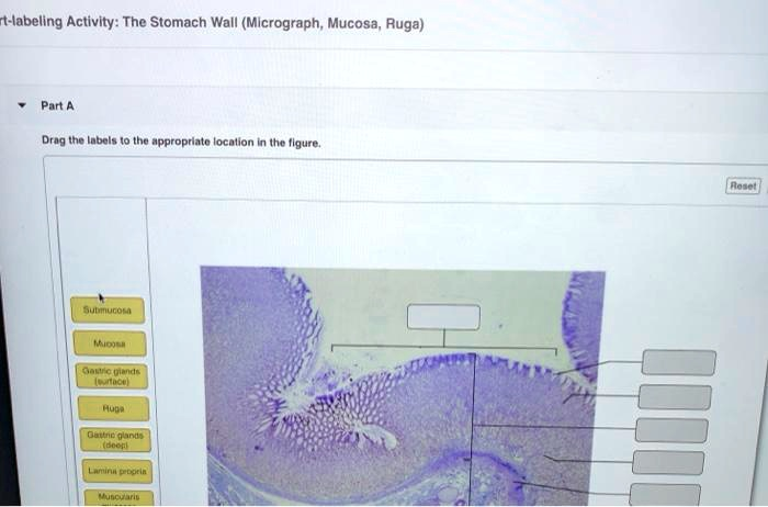
Based on the provided micrograph of the stomach wall, here are the correct labels for the designated boxes.
Correct Labels:
- Top Box: Ruga
- Top Right Box: Mucosa
- Second Right Box: Gastric glands (surface)
- Third Right Box: Gastric glands (deep)
- Fourth Right Box: Lamina propria
- Fifth Right Box: Submucosa
- Bottom Right Box: Muscularis
Explanation of the Stomach Wall Layers
This image provides a microscopic view of the stomach wall, illustrating the specialized layers that enable its digestive functions.
The most prominent structure, labeled at the very top, is a Ruga. These are large, temporary folds in the stomach lining that are visible to the naked eye. Their primary function is to allow the stomach to stretch and expand to accommodate large meals.
The entire innermost layer, indicated by the large bracket, is the Mucosa. This layer is in direct contact with the food we eat and is responsible for secreting protective mucus, hydrochloric acid, and digestive enzymes. The mucosa itself is composed of several sublayers. Its surface is lined with an epithelium that invaginates to form deep pits. These pits lead into the Gastric glands, which are further divided in the diagram. The Gastric glands (surface) refers to the upper portion of these glands, near the opening of the gastric pits. The Gastric glands (deep) points to the lower, main secretory part of the glands. Supporting the epithelium and glands is the Lamina propria, a layer of loose connective tissue containing blood vessels, nerves, and immune cells.
Beneath the mucosa is the Submucosa. This is a layer of dense, irregular connective tissue that provides structural support to the mucosa. It contains larger blood vessels, lymphatic vessels, and a network of nerves called the submucosal plexus, which helps regulate secretions.
The deepest layer shown is the Muscularis, also known as the muscularis externa. This thick layer consists of smooth muscle, typically arranged in three layers in the stomach. Its powerful, coordinated contractions are responsible for the churning and mixing actions that break down food mechanically and propel it into the small intestine