Chloroplast structure Match each image with its label. Photosystem II Granum ATP Chlorophyll a Phospholipid H?O 1/2 NH2 N HO CH HO CH2 Adenine Triphosphate (3 phosphate groups) OH OH Ribose Adenosine (adenine + ribose)
The Correct Answer and Explanation is:
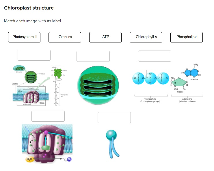
Here are the correct matches for the images:
- Image 1 (Top left diagram with chemical structure): Chlorophyll a
- Image 2 (Top middle 3D chloroplast): Granum
- Image 3 (Top right molecular structure): ATP
- Image 4 (Bottom left protein complex): Photosystem II
- Image 5 (Bottom middle head and tails molecule): Phospholipid
Explanation
This matching exercise identifies key molecules and structures involved in photosynthesis within a chloroplast. Each image represents a specific component essential to this process.
The image in the top left corresponds to Chlorophyll a. This diagram shows the complex chemical structure of the primary photosynthetic pigment. It features a porphyrin ring, which absorbs light energy, and a long hydrocarbon tail that anchors the molecule within the thylakoid membrane. The larger diagram points from a photosystem directly to this molecular structure, indicating its role as a light-harvesting pigment.
The image in the top middle is matched with Granum. This image shows a three dimensional model of a chloroplast. Inside the chloroplast are prominent stacks of flattened, disk like sacs. Each individual stack is called a granum (the plural is grana), and the individual sacs are called thylakoids. The light dependent reactions of photosynthesis occur within the thylakoid membranes that make up the grana.
The image on the top right is the molecular structure of ATP, or Adenosine Triphosphate. The diagram clearly labels its three main components: adenine (a nitrogenous base), ribose (a five carbon sugar), and a chain of three phosphate groups. ATP is the main energy carrying molecule in all living cells, and it is produced during the light dependent reactions of photosynthesis.
The image on the bottom left illustrates Photosystem II. This diagram shows a protein complex embedded in a membrane. It specifically depicts the process of photolysis, where light energy is used to split a water molecule (H₂O) into electrons, protons, and oxygen (½ O₂). This initial light capturing event and the splitting of water are the defining functions of Photosystem II.
Finally, the simple diagram on the bottom middle represents a Phospholipid. This is the classic depiction of a molecule with a hydrophilic (water attracting) head and two hydrophobic (water repelling) tails. Phospholipids are the fundamental building blocks of all biological membranes, including the thylakoid membranes where chlorophyll and the photosystems are located