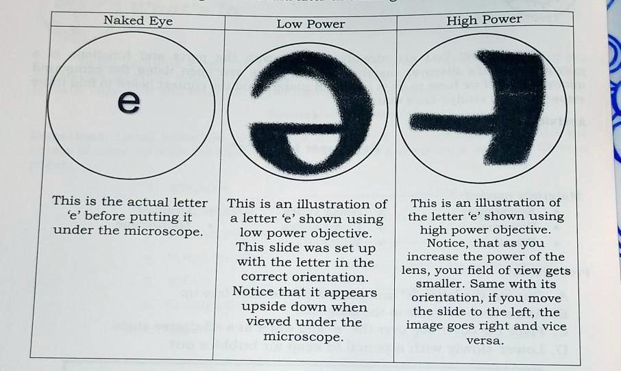
Naked Eye Low Power High Power e This is the actual letter ‘e’ before putting it under the microscope. This is an illustration of a letter ‘e’ shown using low power objective. This slide was set up with the letter in the correct orientation. Notice that it appears upside down when viewed under the microscope. This is an illustration of the letter ‘e’ shown using high power objective. Notice, that as you increase the power of the lens, your field of view gets smaller. Same with its orientation, if you move the slide to the left, the image goes right and vice versa.
The Correct Answer and Explanation is:
The provided image illustrates the fundamental principles of how a compound light microscope alters the image of a specimen. The key takeaways are that the microscope inverts the image both vertically and horizontally, and as magnification increases, the field of view decreases.
Explanation
The diagram effectively demonstrates the optical characteristics of a standard compound microscope by showing the letter ‘e’ as it appears to the naked eye, under low power magnification, and under high power magnification.
First, the most noticeable effect is image inversion. When the letter ‘e’ is placed on the slide in its normal, readable orientation, it appears upside down and reversed when viewed through the eyepiece. This happens because a compound microscope uses a series of convex lenses, specifically the objective lens and the ocular lens (eyepiece). The objective lens produces a real, inverted, and magnified image of the specimen. The eyepiece then further magnifies this already inverted image, presenting the viewer with a final virtual image that is flipped both top to bottom and left to right. This is why the correctly oriented ‘e’ looks like an inverted and mirrored symbol under the low power objective.
Second, the diagram shows the inverse relationship between magnification and the field of view. The field of view is the circular area visible through the microscope. Under the low power objective, the entire letter ‘e’ is visible. However, when switching to the high power objective, the magnification increases significantly. This increased magnification zooms in on a much smaller portion of the specimen. Consequently, the field of view becomes smaller, and only a part of the letter ‘e’, specifically its central crossbar and a section of its curve, can be seen. This illustrates the trade off in microscopy: gaining more detail through higher magnification means losing the broader context of the surrounding area.
Finally, the text points out another consequence of image inversion, which is the apparent reversal of movement. If you physically move the microscope slide to the left, the image you see through the eyepiece will appear to move to the right. Similarly, moving the slide up will cause the image to appear to move down. This occurs because of the complete inversion of the image, and it is a crucial concept for users to grasp in order to navigate a slide and center a specimen effectively.