Art-labeling Activity: Figure 30.2b (1 of 3) Part A Drag the appropriate labels to their respective targets. Right ventricle Mitral valve Left atrium Aortic valve Right atrium Pulmonary valve Tricuspid valve Left ventricle Submit Request Answer Provide Feedback Reset Help
The Correct Answer and Explanation is:
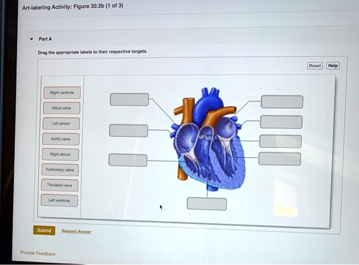
Based on the provided diagram of the human heart, here are the correct labels for each targeted structure:
- Top-left box: Pulmonary valve
- Second box down on the left: Right atrium
- Third box down on the left: Tricuspid valve
- Bottom-left box: Right ventricle
- Top-right box: Aortic valve
- Second box down on the right: Left atrium
- Third box down on the right: Mitral valve
- Bottom-right box: Left ventricle
Explanation of the Heart’s Anatomy and Blood Flow
The provided diagram illustrates the essential structures of the human heart, a four-chambered organ responsible for circulating blood throughout the body via the pulmonary and systemic circuits.
The right side of the heart handles deoxygenated blood. The Right atrium, the upper-right chamber, serves as the initial receiving point for blood returning from the body. This blood then travels through the Tricuspid valve, which ensures one-way flow, into the Right ventricle. The right ventricle is the lower-right chamber that contracts to pump this deoxygenated blood past the Pulmonary valve and into the pulmonary artery, which leads to the lungs for oxygenation.
After gas exchange in the lungs, the newly oxygenated blood returns to the left side of the heart. It enters the Left atrium, the upper-left chamber. From here, the blood flows through the Mitral valve, also known as the bicuspid valve, into the powerful Left ventricle. The left ventricle has a much thicker muscular wall than the right because it must generate enough force to pump blood to the entire body. When the left ventricle contracts, it forces oxygen-rich blood through the Aortic valve and into the aorta, the body’s main artery.
The four valves identified, the tricuspid, pulmonary, mitral, and aortic, are critical for maintaining proper blood flow. They function as one-way gates, opening and closing in a precise sequence with each heartbeat to prevent the backward flow of blood. This coordinated action of the atria, ventricles, and valves ensures an efficient and continuous supply of oxygen and nutrients to all body tissues while removing metabolic waste.