Art-labeling Activity: Internal Anatomy of the Heart (Heart Wall and the Pericardium) Part A Drag the labels to the appropriate location in the figure.
The Correct Answer and Explanation is:
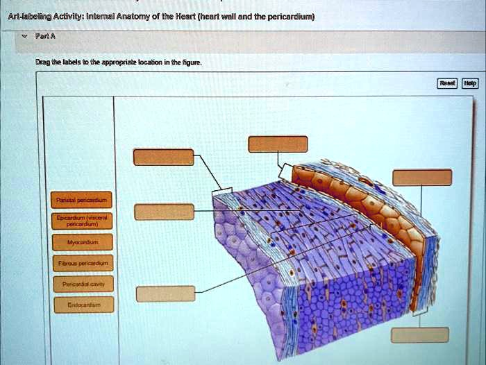
Based on the anatomical structures shown in the diagram, here are the correct labels for the heart wall and the pericardium, matched to their positions:
- Top Right Box: Fibrous pericardium
- Middle Right Box: Parietal pericardium
- Top Left Box: Pericardial cavity
- Bottom Right Box: Epicardium (visceral pericardium)
- Middle Left Box: Myocardium
- Bottom Left Box: Endocardium
Explanation
The provided diagram illustrates a detailed cross section of the layers that make up the heart wall and the surrounding protective sac, the pericardium. Understanding these structures is key to appreciating how the heart is protected and how it functions as a pump.
The heart is enclosed within a double walled sac called the pericardium. The outermost layer, identified as the Fibrous Pericardium, is a tough, dense connective tissue layer. Its primary roles are to protect the heart, anchor it to surrounding structures like the diaphragm and great vessels, and prevent the heart from overfilling with blood.
Deep to this fibrous layer is the two layered serous pericardium. The outer layer of this serous membrane, which lines the internal surface of the fibrous pericardium, is the Parietal Pericardium. The inner layer, which covers the external surface of the heart organ, is the Epicardium, also known as the visceral pericardium. The narrow potential space between these two serous layers is the Pericardial Cavity. This cavity contains a thin film of serous fluid that acts as a lubricant, allowing the heart to beat in a nearly frictionless environment and reducing wear and tear.
The wall of the heart itself consists of three distinct layers. The outermost layer is the Epicardium, as previously mentioned. The middle and by far the thickest layer is the Myocardium. This layer is composed of specialized cardiac muscle tissue and forms the bulk of the heart. The myocardium is the contractile layer responsible for the powerful pumping action that circulates blood throughout the body. The innermost layer, which lines the heart chambers and covers the heart valves, is the Endocardium. It is a thin sheet of simple squamous epithelium that provides a smooth surface to minimize friction as blood flows through the heart. This layer is continuous with the endothelium lining the blood vessels connected to the heart.