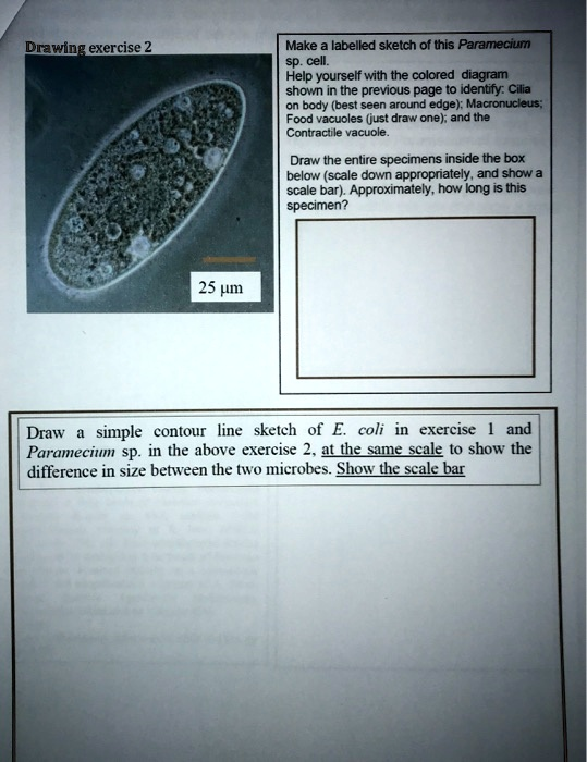
Drawing exercise 2 Make a labelled sketch of this Paramecium sp. cell. Help yourself with the colored diagram shown on the previous page to identify: Cilia body (best seen around the edge), Macronucleus, Food vacuoles (just draw one), and the Contractile vacuole. Draw the entire specimen inside the box below (scale down appropriately, and show a scale bar): Approximately, how long is this specimen? 25 μm Draw a simple contour line sketch of E. coli in exercise and Paramecium sp. in the above exercise 2. Use the same scale to show the difference in size between the two microbes. Show the scale bar.
The Correct Answer and Explanation is:
To draw a labeled sketch of Paramecium sp., let’s break it down and include all the required features:
1. Cilia body
- Paramecia have a dense covering of cilia around their body. These cilia are short, hair-like projections that help the organism move and feed by creating water currents.
- Location: The cilia body is best seen around the edge of the cell.
2. Macronucleus
- Paramecium have two types of nuclei: the macronucleus and the micronucleus. The macronucleus controls the non-reproductive functions of the cell.
- Location: It’s usually large, oval, and located centrally within the cell.
3. Food Vacuoles
- Food vacuoles are the storage structures where food particles are digested.
- Location: They’re scattered throughout the cytoplasm. You can draw one for this sketch.
4. Contractile Vacuole
- This structure helps the Paramecium maintain osmotic balance by expelling excess water.
- Location: Usually located near the center or towards the posterior end of the cell.
Sketch & Scale
- The Paramecium sp. cell is typically about 25 µm long. You can scale this down for your diagram, showing the major components as described above, ensuring clarity and size proportionate to the actual length.
E. coli vs Paramecium size comparison
E. coli is much smaller than Paramecium. On average, E. coli is about 2 µm in length. Paramecium, on the other hand, is roughly 25 µm in length, making it about 10 times larger than E. coli.
For a contour line sketch:
- Draw E. coli as a small rod-shaped organism.
- For Paramecium, use its characteristic elongated shape with a somewhat oval contour and cilia around its perimeter.
Scale Bar
- If you’re scaling your diagrams down proportionately, the E. coli sketch will be much smaller than the Paramecium sketch.
- You can use a 1 µm scale bar for E. coli and a 10 µm scale bar for Paramecium to show the size difference. These will highlight the size contrast clearly.
In your sketch, make sure both organisms are drawn with appropriate labels (Cilia body, Macronucleus, Food vacuoles, and Contractile vacuole for Paramecium) and use the scale bars as indicated.